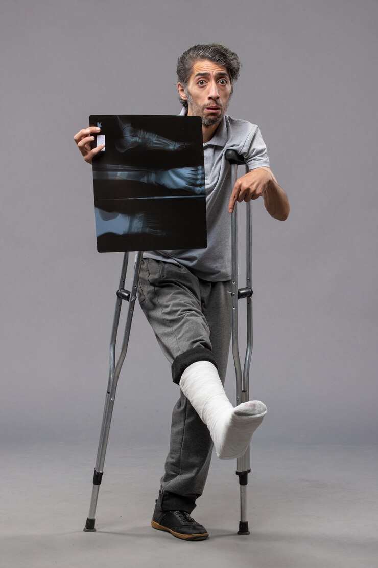
As healthcare professionals, we are often tasked with diagnosing and managing injuries, particularly in primary care settings. One of the most critical aspects of injury management is interpreting X-rays effectively. However, a key component that can enhance our X-ray interpretation skills lies in understanding the mechanism of injury — the story behind how the injury occurred. By taking a thorough patient history, we can significantly improve our ability to identify and interpret injuries correctly, leading to better outcomes for our patients.
Understanding the Mechanism of Injury
The mechanism of injury refers to the way in which an injury occurs. It involves considering the type of force, its direction, the area affected, and the patient’s activity at the time of the injury. When we understand the mechanism, we are better equipped to anticipate the types of fractures or injuries that might be seen on an X-ray.
For example, a fall from height is more likely to result in certain types of fractures, such as a distal radius fracture, while a twisting injury may cause ligament damage or fractures that are more complex. A direct blow to a body part may lead to compression fractures or contusions. Knowing these patterns allows us to focus on the relevant areas during X-ray interpretation and avoid unnecessary or missed diagnoses.
Why Patient Histories Matter in X-ray Interpretation
Patient history is invaluable because it provides context to the images. An X-ray may show a fracture, but without understanding how the injury occurred, it’s difficult to know whether it’s a simple fracture, a more complex injury, or one that requires urgent intervention. For example, a high-energy mechanism of injury such as a car accident may indicate the possibility of multiple fractures or internal injuries that might not be immediately apparent on the X-ray. On the other hand, a low-energy fall in an elderly patient may suggest a simple fracture, such as a hip fracture or wrist fracture.
By carefully asking the right questions about the injury — for example, how the injury occurred, what the patient was doing at the time, and whether there was any immediate pain, swelling, or deformity — we can often determine what to look for when reviewing an X-ray. This practice not only enhances the accuracy of our diagnoses but also allows us to make more informed decisions about next steps in patient care.
Common Injury Mechanisms and X-ray Implications
- Falls from Height
These injuries are often associated with fractures to the lower extremities, spine, and upper limbs. A thorough history is essential to determine the force involved and to rule out associated injuries. For example, a fall onto an outstretched hand can result in a Colles’ fracture or scaphoid fracture. - Sports Injuries
Sports-related injuries can range from simple soft tissue damage to complex fractures. A quick understanding of the mechanism can help identify common patterns, such as ligament tears or fractures due to twisting or impact forces. For instance, a contact sport injury may result in a knee ligament injury, while a twisting motion could lead to ankle fractures. - Direct Blows
Direct trauma, such as a blow to the face or limbs, can result in fractures like nasal bone fractures or fractures of the tibia and fibula. Understanding whether the injury was caused by a blunt or penetrating force will help clinicians recognize fractures and other internal damage that may not be visible initially. - Motor Vehicle Accidents (MVAs)
MVAs often involve high-energy forces that can lead to serious injuries, including pelvic fractures, fractures of the spine, and internal injuries. Understanding the direction and impact of the collision helps in assessing the potential injury severity and guides appropriate X-ray protocols.
Enhancing Your Skills: X-ray Interpretation Courses
Effective X-ray interpretation is a skill that requires practice and continual learning, especially in the context of minor injuries. Healthcare professionals can benefit from formal training to refine their diagnostic abilities and ensure they are correctly identifying and managing injuries.
If you are looking to enhance your X-ray interpretation skills, consider the following Practitioner Development UK courses:
- X-ray Interpretation of Minor Injuries (Includes Red Dot)
This course is designed to improve the diagnostic accuracy of healthcare professionals when interpreting X-rays of minor injuries. The addition of the Red Dot system helps practitioners identify serious injuries that require immediate attention. This course will deepen your understanding of common injury patterns and provide practical knowledge for interpreting X-rays with confidence. - Minor Injury Essentials – Face-to-Face Accredited by the RCN Centre for Professional Accreditation
This comprehensive, face-to-face course offers in-depth training for primary care healthcare professionals in managing minor injuries. Accredited by the RCN Centre for Professional Accreditation, this course combines clinical knowledge with practical skills, giving attendees the tools needed to assess, diagnose, and treat minor injuries effectively.
By enrolling in both the X-ray Interpretation of Minor Injuries and Minor Injury Essentials courses, healthcare professionals can gain a firm grounding in managing minor injuries. These courses provide a comprehensive understanding of injury mechanisms and an essential framework for interpreting X-rays accurately. Combined, they offer a holistic approach to assessing and managing minor injuries, helping healthcare professionals make more informed decisions in their daily practice.
Conclusion
Incorporating an understanding of injury mechanisms into your clinical practice can greatly enhance your ability to interpret X-rays accurately. By gathering a detailed patient history and considering the forces at play, you can ensure that you are not only identifying injuries correctly but also managing them appropriately. Through courses such as the ones mentioned above, you can refine these essential skills and provide better care for your patients. Stay proactive in your professional development and enhance your ability to deliver accurate diagnoses through thoughtful X-ray interpretation.
References
Leach, B. (2023). A comprehensive guide to the management of minor injuries in primary care. British Journal of Primary Care, 28(3), 155-161.
Smith, A. & Williams, H. (2022). X-ray interpretation in the primary care setting: An overview for healthcare professionals. Journal of Clinical Radiology, 56(7), 22-29.


